Staff profile
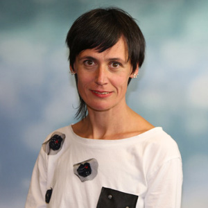
Dr Margarita Staykova
Associate Professor

| Affiliation | Telephone |
|---|---|
| Associate Professor in the Department of Physics | +44 (0) 191 33 43598 |
| Associate Fellow in the Institute of Advanced Study | |
| Associate Professor in the Biophysical Sciences Institute | |
| Biophysical Sciences Institute Executive Board in the Biophysical Sciences Institute | |
| Associate Professor in the Durham CELLS (Centre for Ethics and Law in the Life Sciences) | |
| Co-Director (Science) in the Institute of Advanced Study | +44 (0) 191 33 43598 |
Biography
Margarita Staykova is Associate Professor at the Department of Physics, Durham University, where she leads a research group in Biophysics and Soft Condensed Matter. She is Director of Science at the Institute of Advanced Study, and Executive Board Member at the Biophysics Science Institute. Prior to joining Durham, Margarita has worked in the Department of Mechanical Engineering at Princeton University (2009-2013) and at the Max Planck Institute of Colloids and Interfaces, Germany (2007-2009). She has a PhD in Biophysics from Rostock University, Germany (2006) and MSc in Molecular Biology from Sofia University, Bulgaria.
Group
Margarita leads a biophyiscs group that combines experiments on membrane model systems and on living cells, and uses those to design bio-hybrid systems with life like properties. We are interested in the ethical, technological and social implications of our research and explore them through close collaborations with science and technology scholars, anthropologists and artists.
Margarita welcomes inquiries from postgraduates in any aspect of membrane/cell biophysics and bio-engineering, and is always open to new collaborations with scientists and stakeholders across various fields.
Biophysics
The cell interface is a multi-layered ensemble, with the plasma membrane in the middle, a contractile actin cortex on the inner side, and an extracellular matrix, cell wall or neighbour cell membrane on the outer side. To understand how the coupling between these structurally and mechanically different layers shape the surface functionality of cells, we develop synthetic cells and model interfaces using tools from soft matter and microfluidics, and subject them to mechanical and electrical manipulation.
For example, by coupling lipid membranes to elastic substrates we were able to demonstrate how cells regulate passively their surface area under mechanical perturbation, how mechanical stretch and compression regulate membrane protein binding (Roux et al.) and cholesterol content (Rahimi et al; Miller et al.), and how membranes open and close pores in controllable manner (Goodband et al.). In a more recent study (Dinet et al), we provided the experimental and physical understanding of how hydraulic fracturing can pattern cell-cell adhesions and facilitate biological processes such as lumen formation.
With the help of such experiments we aim to understand the mechano-sensitive organisation of biological interfaces, and reconstitute it in biomimetic and bio-hybrid systems of academic and industrial interest. We work with industrial partners such as GSK, Procter&Gamble, and Mondelēz.
Biohybrid futures
In our Material Imagination project (https://materialimagination.org), we study the interactions of microbes with lipid membranes in order to understand the principles and mechanisms of cellular pathogenesis and endosymbiosis, and to design biohybrid systems with the life-like abilities to move, adapt and propagate. In a close collaboration with the social scientist Tiago Moreira, the artist Alexandra Carr and fellows of the Institute of Advances Study, we explore the social and future dimensions of creating and living with biohybrid technologies, that extend beyond the immediate scope of scientific explorations (Moreira et al.). For more information, please take a look at our video, commissioned by the Royal Society (https://www.youtube.com/watch?v=sK32DnuMnOU)
Connect
Margarita has sponsored a number of Visiting IAS Fellows to Durham University:
2023 Professor Elisabeth A. Povinelli, Department of Anthropology, Columbia University, USA
2023 Professor David Kneas, Department of Anthropology, University of South Carolina, USA
2020 Professor Wilson Poon, Department of Physics, University of Edinburgh, UK
2020 Professor Torben E Jensen, Techno-Anthropology Research group, Aalborg University, Denmark.
2020 Alexandra Carr, artist
2020 Professor Laura Forlano, College of Arts, Media, and Design, Northeastern University, USA
Research interests
- Biophysics
- Biological membranes
- Lipid interfaces
- Synthetic cells
- Living materials
- Responsible research and Innovation
- Science and Technology Studies
Esteem Indicators
- 2023: Director for Science - Institute of Advanced Study, Durham University :
Publications
Chapter in book
- Biophysical insights from supported lipid patchesMiller, E., Stubbington, L., Dinet, C., & Staykova, M. (2019). Biophysical insights from supported lipid patches. In A. Iglič, M. Rappolt, & A. J. García-Sáez (Eds.), Advances in Biomembranes and Lipid Self-Assembly. (pp. 23-48). Elsevier. https://doi.org/10.1016/bs.abl.2019.01.004
Journal Article
- Lipid bilayer fracture under uniaxial stretchGoodband, R. J., & Staykova, M. (2025). Lipid bilayer fracture under uniaxial stretch. Soft Matter, 21(9), 1669-1675. https://doi.org/10.1039/d4sm01410c
- Co-developing materials in the metamorphic zone: extending bacteriocentricityMoreira, T., & Staykova, M. (2024). Co-developing materials in the metamorphic zone: extending bacteriocentricity. Science, Technology, & Human Values. Advance online publication. https://doi.org/10.1177/01622439241296898
- Engaging publics in imagining the future of engineered living materialsMoreira, T., Marshall, J., & Staykova, M. (2023). Engaging publics in imagining the future of engineered living materials. Matter, 6(8), 2467-2470. https://doi.org/10.1016/j.matt.2023.05.002
- Patterning and dynamics of membrane adhesion under hydraulic stressDinet, C., Torres-Sánchez, A., Lanfranco, R., Di Michele, L., Arroyo, M., & Staykova, M. (2023). Patterning and dynamics of membrane adhesion under hydraulic stress. Nature Communications, 14(1), Article 7445. https://doi.org/10.1038/s41467-023-43246-7
- Scratching beyond the surface — minimal actin assemblies as tools to elucidate mechanical reinforcement and shape changeAufderhorst-Roberts, A., & Staykova, M. (2022). Scratching beyond the surface — minimal actin assemblies as tools to elucidate mechanical reinforcement and shape change. Emerging Topics in Life Sciences, 6(6), 583-592. https://doi.org/10.1042/etls20220052
- Comparative Study of Lipid- and Polymer-Supported Membranes Obtained by Vesicle FusionGoodband, R. J., Bain, C. D., & Staykova, M. (2022). Comparative Study of Lipid- and Polymer-Supported Membranes Obtained by Vesicle Fusion. Langmuir, 38(18), 5674-5681. https://doi.org/10.1021/acs.langmuir.2c00266
- Encapsulated bacteria deform lipid vesicles into flagellated swimmersLe Nagard, L., Brown, A. T., Dawson, A., Martinez, V. A., Poon, W. C., & Staykova, M. (2022). Encapsulated bacteria deform lipid vesicles into flagellated swimmers. Proceedings of the National Academy of Sciences, 119(34). https://doi.org/10.1073/pnas.2206096119
- Dynamic Mechanochemical feedback between curved membranes and BAR protein self-organizationRoux, A. L., Tozzi, C., Walani, N., Quiroga, X., Zalvidea, D., Trepat, X., Staykova, M., Arroyo, M., & Roca-Cusachs, P. (2021). Dynamic Mechanochemical feedback between curved membranes and BAR protein self-organization. Nature Communications, 12, Article 6550. https://doi.org/10.1038/s41467-021-26591-3
- Substrate-led cholesterol extraction from supported lipid membranesMiller, E., Voitchovsky, K., & Staykova, M. (2018). Substrate-led cholesterol extraction from supported lipid membranes. Nanoscale, 10(34), 16332-16342. https://doi.org/10.1039/c8nr03399d
- Shape Transformations of Lipid Bilayers Following Rapid Cholesterol UptakeRahimi, M., Regan, D., Arroyo, M., Subramaniam, A., Stone, H., & Staykova, M. (2016). Shape Transformations of Lipid Bilayers Following Rapid Cholesterol Uptake. Biophysical Journal, 111(12), 2651-2657. https://doi.org/10.1016/j.bpj.2016.11.016
- Biophysical dissection of schistosome septins: Insights into oligomerization and membrane bindingZeraik, A., Staykova, M., Fontes, M., Nemuraitė, I., Quinlan, R., Araújo, A., & DeMarco, R. (2016). Biophysical dissection of schistosome septins: Insights into oligomerization and membrane binding. Biochimie, 131, 96-105. https://doi.org/10.1016/j.biochi.2016.09.014
- Sticking and sliding of lipid bilayers on deformable substratesStubbington, L., Arroyo, M., & Staykova, M. (2016). Sticking and sliding of lipid bilayers on deformable substrates. Soft Matter, 13, 181-186. https://doi.org/10.1039/c6sm00786d
- Responsible innovation across borders: tensions, paradoxes and possibilitiesMacnaghten, P., Owen, R., Stilgoe, J., Wynne, B., Azevedo, A., de Campos, A., Chilvers, J., Dagnino, R., di Giulio, G., Frow, E., Garvey, B., Groves, C., Hartley, S., Knobel, M., Kobayashi, E., Lehtonnen, M., Lezaun, J., Mello, L., Monteiro, M., … Velho, L. (2014). Responsible innovation across borders: tensions, paradoxes and possibilities. Journal of Responsible Innovation., 1(2), 191-199. https://doi.org/10.1080/23299460.2014.922249
- Confined bilayers passively regulate shape and stressStaykova, M., Arroyo, M., Rahimi, M., & Stone, H. (2013). Confined bilayers passively regulate shape and stress. Physical Review Letters, 110(2), Article 028101. https://doi.org/10.1103/physrevlett.110.028101
- Mechanics of surface area regulation in cells examined with confined lipid membranes.Staykova, M., Holmes, D., Read, C., & Stone, H. (2011). Mechanics of surface area regulation in cells examined with confined lipid membranes. Proceedings of the National Academy of Sciences, 108(22). https://doi.org/10.1073/pnas.1102358108
- Wrinkling and electroporation of giant vesicles in the gel phase.Knorr, R., Staykova, M., Gracija, R., & Dimova, R. (2010). Wrinkling and electroporation of giant vesicles in the gel phase. Soft Matter, 6(9), 1990-1996. https://doi.org/10.1039/b925929e
- Vesicles in electric fields: Some novel aspects of membrane behavior.Dimova, R., Bezlyepkina, N., Jordö, M., Knorr, R., Riske, K., Staykova, *M., Vlahovska, P., Yamamoto, T., Yang, P., & Lipowsky, R. (2009). Vesicles in electric fields: Some novel aspects of membrane behavior. Soft Matter, 5(17), 3201-3212. https://doi.org/10.1039/b901963d
- Membrane flow patterns in multicomponent giant vesicles induced by alternating electric fields.Staykova, M., Lipowsky, R., & Dimova, R. (2008). Membrane flow patterns in multicomponent giant vesicles induced by alternating electric fields. Soft Matter, 4(11), 2168-2171. https://doi.org/10.1039/b811876k
- The influence of the molecular structure of lipid membranes on the electric field distribution and energy absorptionSimeonova, M., & Gimsa, J. (2006). The influence of the molecular structure of lipid membranes on the electric field distribution and energy absorption. Bioelectromagnetics, 27(8), 652-666. https://doi.org/10.1002/bem.20259
- Dielectric anisotropy, volume potential anomalies and the persistent Maxwellian equivalent bodySimeonova, M., & Gimsa, J. (2005). Dielectric anisotropy, volume potential anomalies and the persistent Maxwellian equivalent body. Journal of Physics: Condensed Matter, 17(50), 7817-7831. https://doi.org/10.1088/0953-8984/17/50/004
- Estimating the subcellular absorption of electric field energy: equations for an ellipsoidal single shell modelWachner, D., Simeonova, M., & Gimsa, J. (2002). Estimating the subcellular absorption of electric field energy: equations for an ellipsoidal single shell model. Bioelectrochemistry, 56(1-2). https://doi.org/10.1016/s1567-5394%2802%2900020-8
- Cellular absorption of electric field energy: influence of molecular properties of the cytoplasm.Simeonova, M., Wachner, D., & Gimsa, J. (2002). Cellular absorption of electric field energy: influence of molecular properties of the cytoplasm. Bioelectrochemistry, 56(1-2). https://doi.org/10.1016/s1567-5394%2802%2900010-5

