/prod01/prodbucket01/media/durham-university/research-/new-research-and-business/Research-and-Business-Partnerships.png)
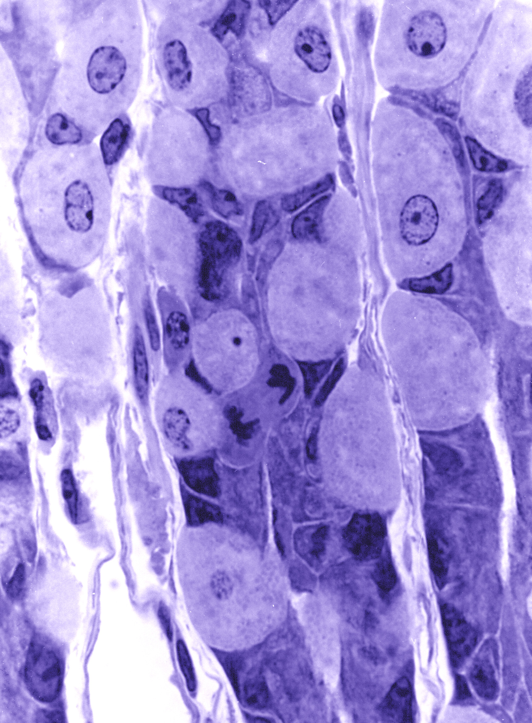
Image above: Microscopy image of the gastric glands of the lining of the stomach, courtesy of Dr R W Banks
Gastric glands of the lining of the stomach
The large, pale cells secrete hydrochloric acid; the smaller, darker cells secrete mucus. One cell near the centre of the image is dividing, having almost completed anaphase when the two sets of daughter chromosomes move apart. The tissue has been chemically fixed, dehydrated, and embedded in epoxy resin. The section was cut with a glass knife and is 1 micrometre thick. It was stained with toluidine blue for light microscopy.