/prod01/prodbucket01/media/durham-university/research-/new-research-and-business/Research-and-Business-Partnerships.png)
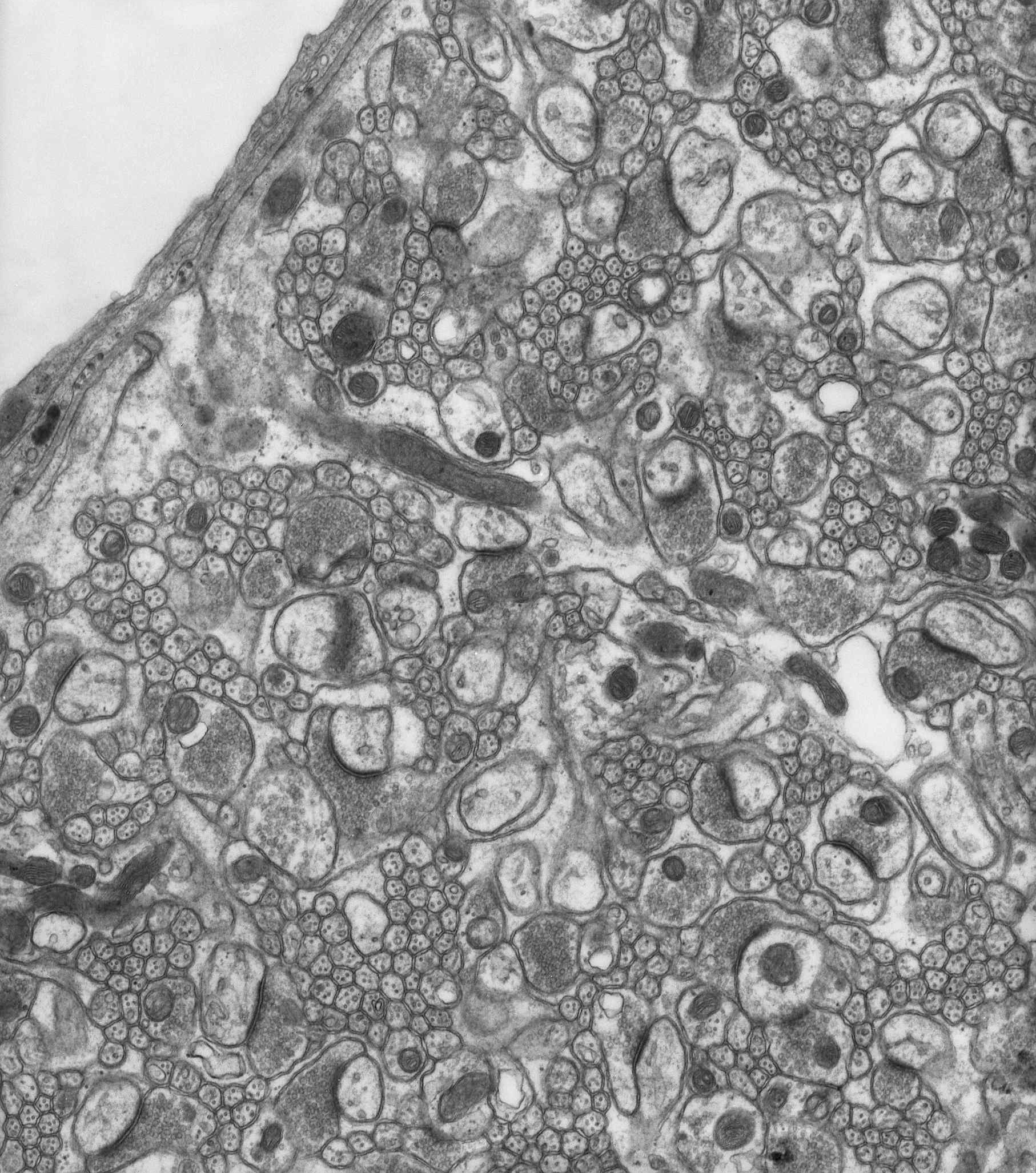
Image above: Electron micrograph image of the outer (molecular) layer of the cerebellar cortex, courtesy of Dr R W Banks.
The outer layer of the cerebellar cortex
An electron micrograph of the outer (molecular) layer of the cerebellar cortex, containing many connexions, or synapses, between nerve cells. The cerebellum is a large part of the brain, and most of its complex functions are automatic. Disease of, or damage to, the cerebellum is often associated with loss of normal motor control. The tissue has been chemically fixed, dehydrated, and embedded in epoxy resin. The section was cut with a glass knife and is about 80 nanometres thick. It was “stained” with lead citrate and uranyl acetate to provide sufficient contrast in the electron microscope.