/prod01/prodbucket01/media/durham-university/research-/new-research-and-business/Research-and-Business-Partnerships.png)
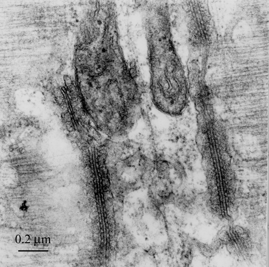
Image above: Electron micrograph image showing internal membranous structures deep within the substance of a skeletal muscle fibre, courtesy of Dr R W Banks.
Structures Deep Within Skeletal Muscle Fibre
An electron micrograph showing internal membranous structures deep within the substance of a skeletal muscle fibre. At the top are a pair of mitochondria that provide energy for muscle contraction. The elongate structures running from top to bottom of the image are called triads because of their three membranous components. The middle of the triad is the transverse (or T-) tubule, which carries the electrical command to contract deep into the muscle fibre. It is connected to sac-like compartments (the sarcoplasmic reticulum) on either side by the row of dark, peg-like structures. When the electrical signal reaches them, the “pegs” open minute channels releasing calcium ions from the sarcoplasmic reticulum, and the calcium ions instruct the contractile proteins of the muscle fibre to begin to contract. The tissue has been chemically fixed, dehydrated, and embedded in epoxy resin. The section was cut with a glass knife and is about 80 nanometres thick. It was “stained” with lead citrate and uranyl acetate to provide sufficient contrast in the electron microscope.