/prod01/prodbucket01/media/durham-university/research-/73895-2-1920X290.jpg)
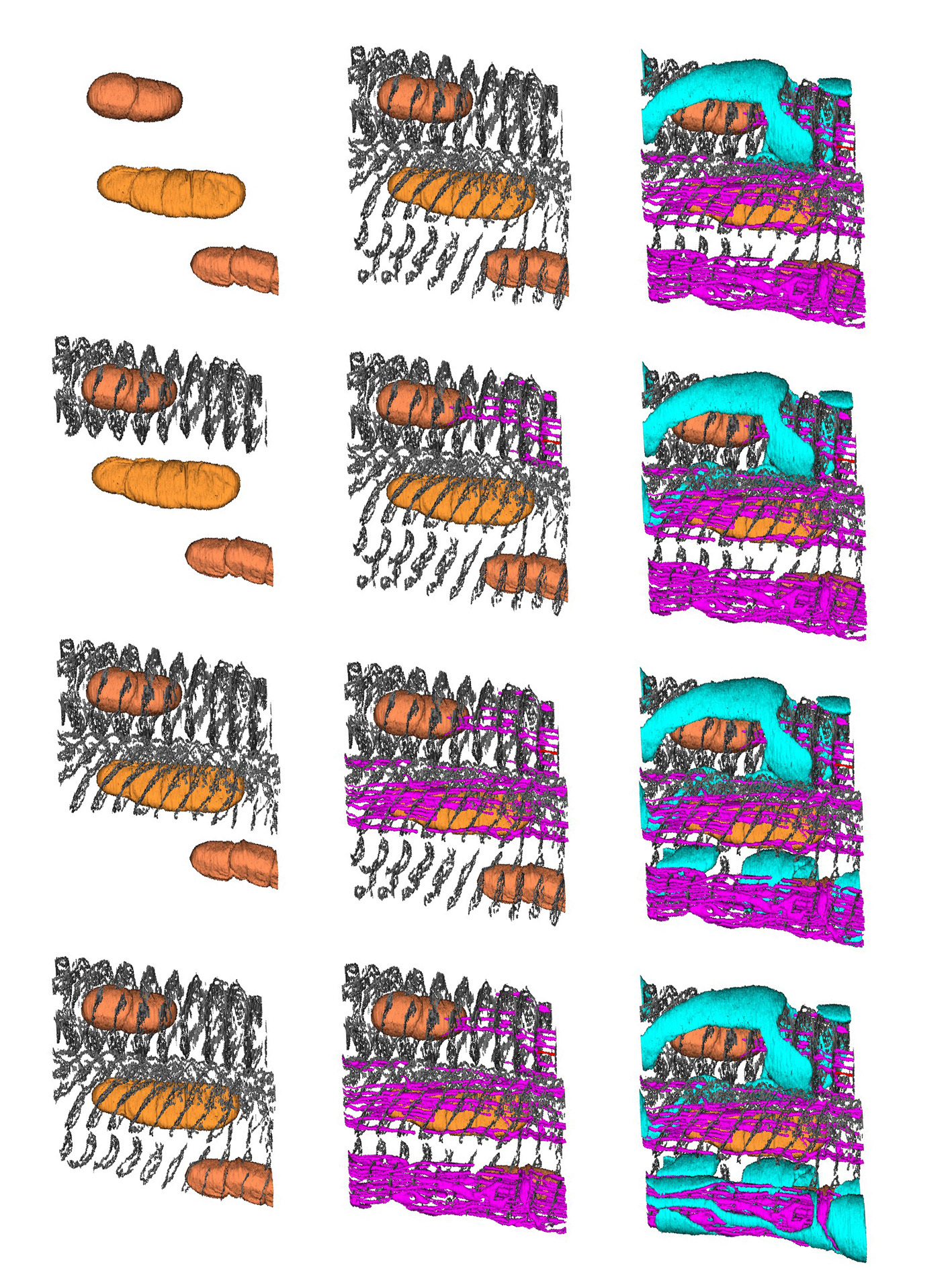
Image above: Serial block-face scanning electron microscopy (SBF-SEM) reconstructed image showing a section of a muscle spindle, courtesy of Dr R W Banks.
Section of a Muscle Spindle
Muscle spindles are sense organs in skeletal muscles that respond to the length and changing length of muscles. They are extremely important in motor control. These images show a virtual reconstruction of a small portion of a muscle spindle using a technique known as serial block-face scanning electron microscopy (SBF-SEM) in which thin slices are repeatedly taken off the face of the block of tissue. A scanning electron micrograph is made of each successive face of the block. This reconstruction was made from 300 serial images, each 70 nanometres apart. Starting at the top left the images progressively include additional features, as follows: the brownish blobs are nuclei in specialised muscle fibres within the spindle; the dark grey lacey things are parts of the contractile structure of the muscle fibres (Z-lines); the purple things are mitochondria in some of the muscle fibres; and the turquoise things are sensory nerve endings.