/prod01/prodbucket01/media/durham-university/research-/new-research-and-business/Research-and-Business-Partnerships.png)
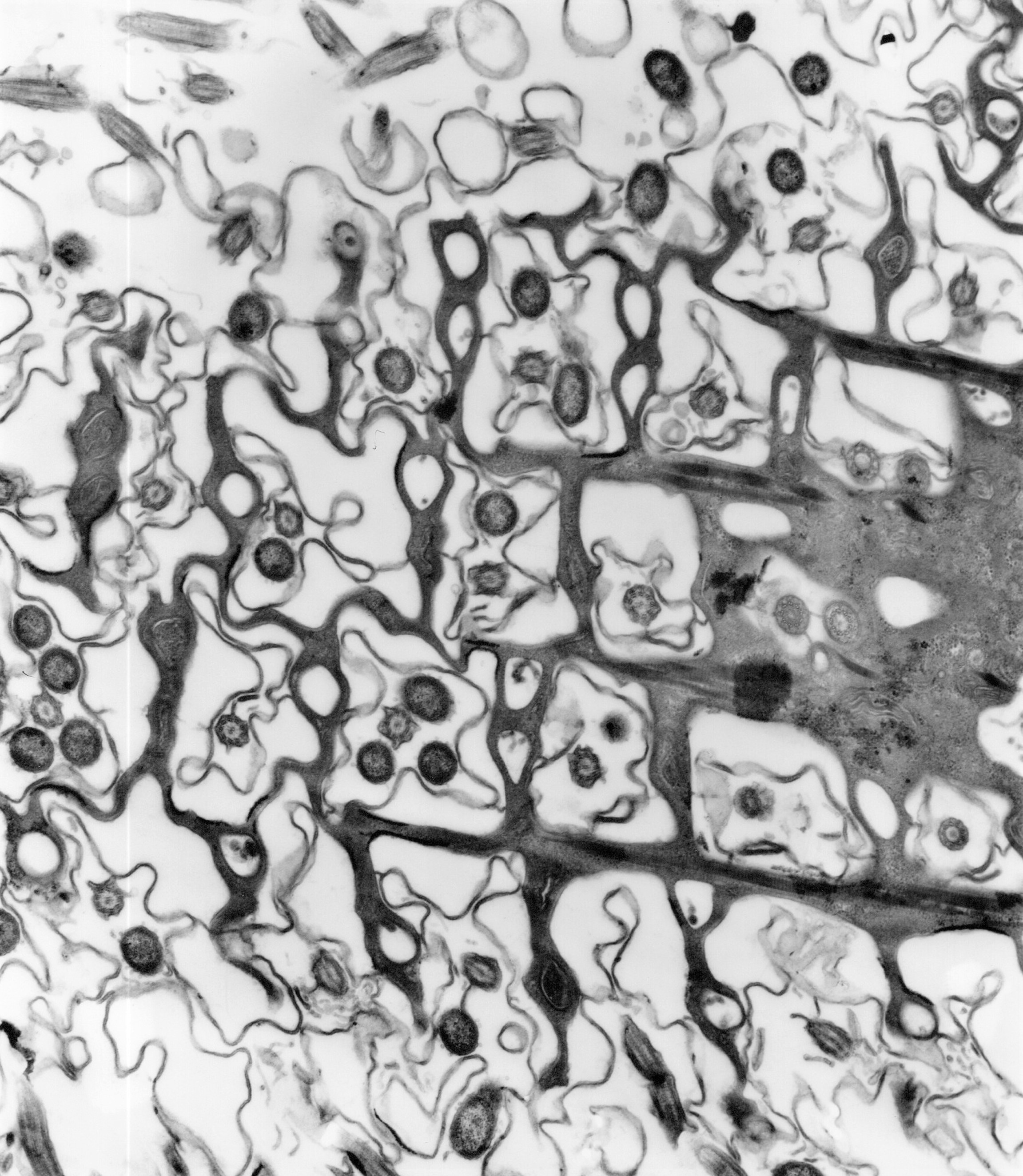
Image above: Electron microscope image of Paramecium, courtesy of Dr R W Banks.
Paramecium
This is an example of a free-living, single-celled organism, known as Paramecium. Paramecium lives in fresh water; it moves and feeds using tiny, hair-like projections from the surface of the cell, called cilia (singular – cilium). The image is of a section of about 80 nanometres thickness passing almost parallel to the cell surface, which is sculpted into squarish pockets with a cilium at the centre of each one. Next to the cilium in many of the pockets are symbiotic bacteria that appear as dark, roughly circular structures. Lead citrate and uranyl acetate “stains” were used to provide contrast in the electron microscope.