/prod01/prodbucket01/media/durham-university/research-/new-research-and-business/Research-and-Business-Partnerships.png)
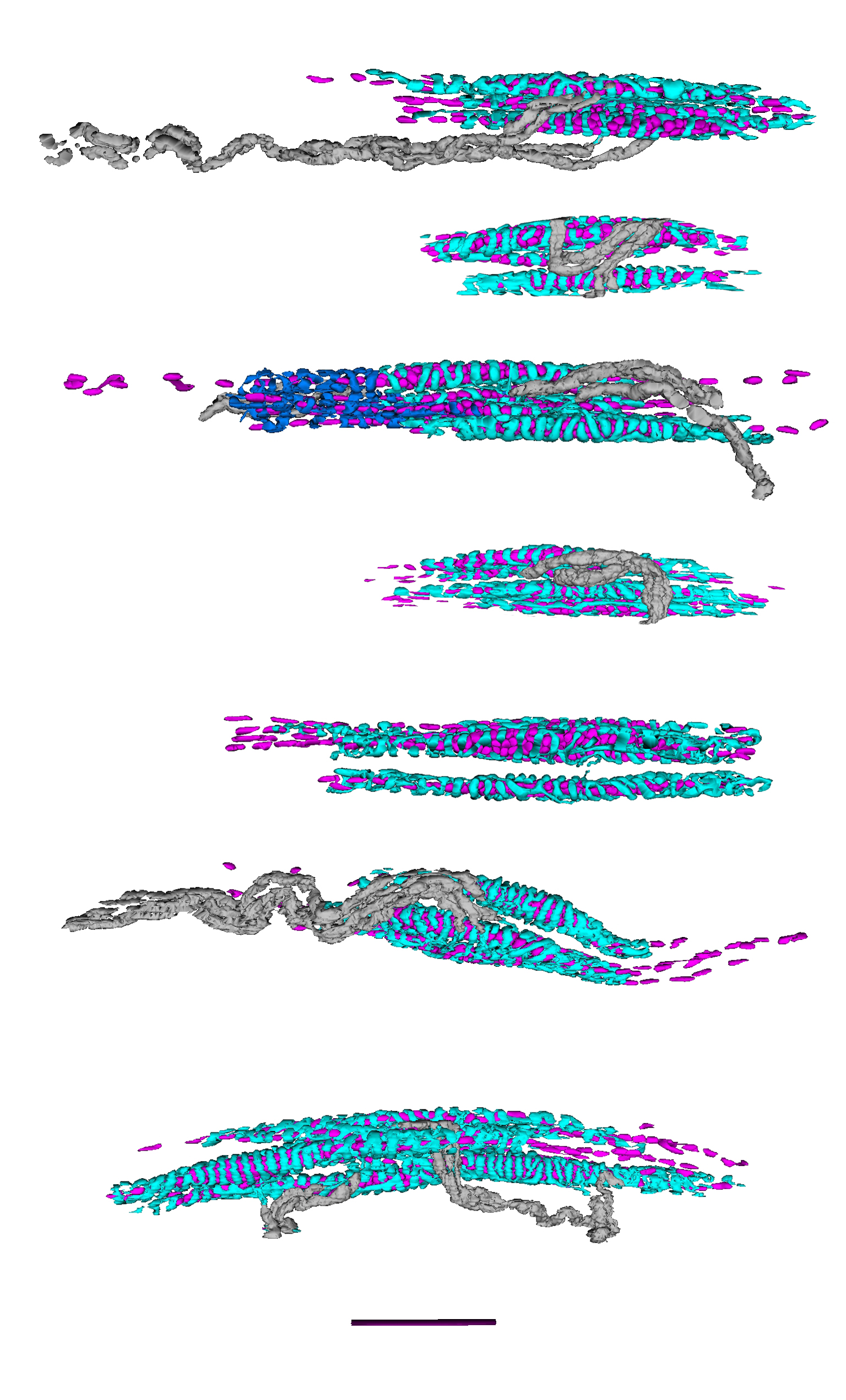
Image above: These images show virtual reconstructions of the main (“primary”) sensory endings of seven spindles, made using serial sections for light microscopy, courtesy of Dr R W Banks.
Sensory Endings of Muscle Spindles
Muscle spindles are sense organs in skeletal muscles that respond to the length and changing length of muscles. They are extremely important in motor control. These images show virtual reconstructions of the main (“primary”) sensory endings of seven spindles, made using serial sections for light microscopy. Each section was I micrometre thick and they were stained with toluidine blue dye. The colours used in the reconstructions are arbitrary: sensory endings, with characteristic annulo-spiral form, are shown in turquoise; they are supplied by myelinated nerve fibres shown in grey. Annulo-spiral endings are wrapped around specialised muscle fibres, only the nuclei of which are shown (in purple).