/prod01/prodbucket01/media/durham-university/research-/new-research-and-business/Research-and-Business-Partnerships.png)
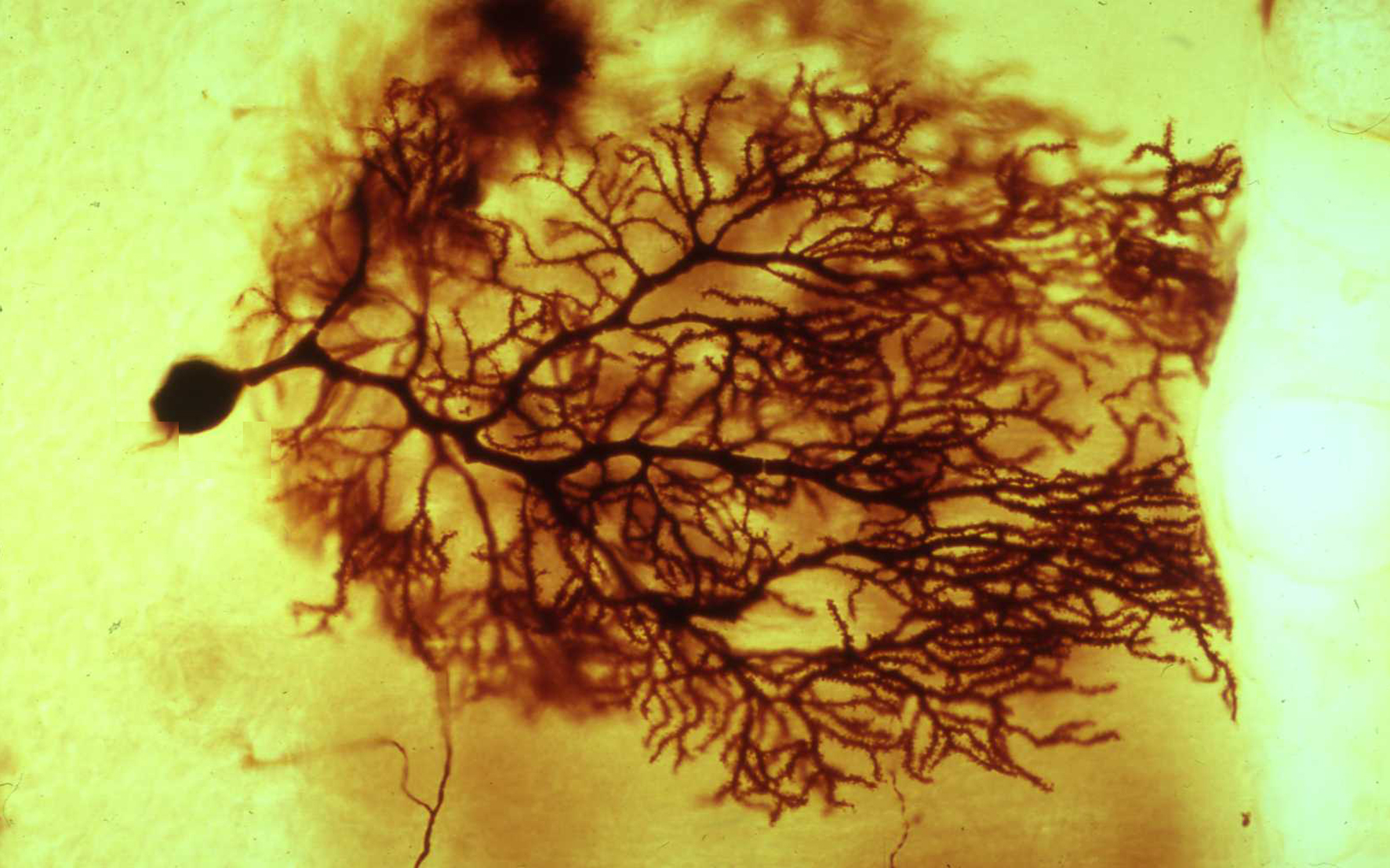
Image above: Microscopy image of a Purkinje cell, one type of nerve cell, or neuron, from the cortex of the cerebellum, courtesy of Dr R W Banks.
A nerve cell, or neuron, from the cortex of the cerebellum
The cerebellum is a large part of the brain, and most of its complex functions are automatic. Disease of, or damage to, the cerebellum is often associated with loss of normal motor control. The tissue has been chemically fixed, dehydrated, and embedded in epoxy resin. The section was cut with a steel knife and is about 100 micrometres thick. There are many hundreds of different types of neuron in the nervous system, of which this, the Purkinje cell, is just one example. Its body, or soma, contains the cell’s nucleus and is the dark blob near the botom of the image. The very extensive, tree-like branches arising from the soma, the dendrites, receive synaptic input from thousands of other neurons. The dendrites and soma integrate the information to produce an output in the form of nerve impulses that travel away from the soma along the axon, the start of which is just visible on the lower right of the soma. The Purkinje cell has been visualised by the Golgi method that more or less randomly stains a few neurons with a silver chromate precipitate, leaving the majority unstained.