Brightfield & Widefield Fluorescence Microscopy
Brightfield and widefield fluorescence microscopes offer the simplest and most widely used methods in light microscopy.
Brightfield & Widefield Fluorescence Microscope Systems
GE Healthcare OMX V4.0
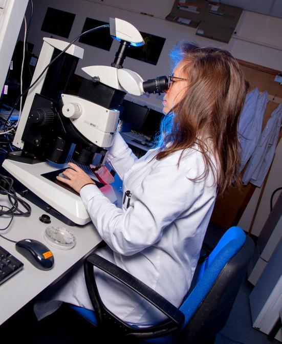
Leica M165FC Stereo
This fluorescence stereo microscope combines high resolution with large depth of field and large zoom range and is ideally suited to tasks which require imaging of whole organisms and manipulation of these under fluorescence illumination. The system can capture every aspect of an organism over a wide magnification range (7.3x - 120x), down to the tiniest details, resolving structures down to a size of 551nm allowing the user to obtain overview and detailed image acquisition in 1 step. The microscope has encoding of the focus, zoom, filters, and iris diaphragm and this configuration and optical data can be read out at the computer at any time and associated with the image as meta data.

General Specification
Stereo microscope
Fluorescence capabilities - GFP2 & DS Red filter cubes
Leica camera
Objective Lenses
- Leica PlanApo 1.0x
- Leica PlanApo 2.0x


/prod01/prodbucket01/media/durham-university/departments-/biosciences/83453-1-1595X1594.jpg)
/prod01/prodbucket01/media/durham-university/departments-/biosciences/infrastructure-/bioimaging/IMG_0298.jpg)
/prod01/prodbucket01/media/durham-university/departments-/biosciences/infrastructure-/bioimaging/12464.jpg)
/prod01/prodbucket01/media/durham-university/departments-/biosciences/infrastructure-/bioimaging/IMG_0347.jpg)
/prod01/prodbucket01/media/durham-university/departments-/biosciences/infrastructure-/bioimaging/Bioimaging-rectangle.png)
/prod01/prodbucket01/media/durham-university/departments-/biosciences/infrastructure-/bioimaging/IMG_0368-1.jpg)
/prod01/prodbucket01/media/durham-university/departments-/biosciences/infrastructure-/bioimaging/Cell-Observer.jpg)
/prod01/prodbucket01/media/durham-university/departments-/biosciences/infrastructure-/bioimaging/OMX.jpg)